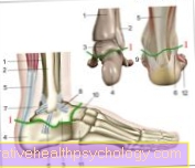
Figur revet ligament
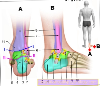
- Fremre fibula -
Ankelbånd -
Lig. Fibulotalare anterius - Fibula-calcaneus
Bånd -
Calcaneofibular ligament - Posterior fibula
Ankelbånd -
Posterior fibulotalar ligament - Hælben - calcaneus
- Ankelben - Talus
- Ydre ankel -
(= Fibulaknogel)
Lateral malleolus - Fibula - fibula
- Shin - tibia
- Cuboid knogel -
Os cuboideum - Scaphoid (af foden) -
Navikulære knogler - Indvendig ankel -
(= Shin bone) -
Medial malleolus
I - I - Øvre ankel
(Hængsel linje blå) -
Articulatio talocruralis
II - II - Under ankel
(Hængsel linje lilla) -
Articulatiotalocalcaneonavicularis
Du kan finde en oversigt over alle Dr-Gumpert-billeder på: medicinske illustrationer
Relaterede billeder

Illustration
fod bøjes over
Illustration
Øvre ankel
Illustration
Under ankel

Illustration
Smerter i læggen

Illustration
forstuvning

Illustration
fibula

Figur muskler
gastrocnemius
(Lægmuskel)